 Heel Pain & Plantar Fasciitis

We currently run specialist plantar fasciitis and chronic heel pain
clinics from Sheffield & Nottingham. For specialist heel pain appointments please
call 0845 2242576 please state that you require a heel pain
assessment. All three heel pain clinics are located in central
locations only minutes away from train stations by taxi.
Since 1999 we have developed a wealth of
experience in treating people from all over the country with
persisting heel pain. We receive referrals nation wide from a
wide range of health professionals including GPs,
Physiotherapists, Orthopaedic Consultants, Rheumatologists,
Sports Medicine Physicians and other Podiatrists and tennis and
golf coaches.
We specialise in stubborn persisting cases
of heel pain. The average patient we see with heel pain will
typically have suffered symptoms over eighteen months. Patients
will have often had other forms of treatment. Our treatments
enjoy high success rates, with typically 85% of patients being
symptom free or significantly improved* (*defined as, a decrease
in frequency and severity of symptoms by an order of 60-70%)
within the first 6 weeks of treatment. This level of success is
based on experience of treating thousands of patients and
thorough assessment of your condition. Investigations can
include x rays, MRI scanning and in-house ultrasound scanning
and computerised gait analysis (an assessment of how you foot
functions when walking).
The
most common cause of deep pain on the bottom surface of the
heel is Plantar Fasciitis (inflammation of the plantar fascia).
The plantar fascia is a broad band of fibrous tissue which
runs along the bottom surface of the foot, from the heel to
the toes. It is just below the skin and subcutaneous fat.
It helps to secure the arch of the foot. Long standing inflammation
causes the deposition of calcium at the point where the plantar
facia inserts into the heel bone. This can result in the appearance
of a bony heel spur on x-ray. The spur itself is not the source
of the pain. Stubborn heel pain should be evaluated by a Podiatrist.
Plantar fasciitis may also present as pain anywhere along
the sole of the foot, particularly along the arch and just
in front of the heel.
Symptoms
- Sharp pain often localised to the bottom and/or inside
margin of the heel
- Pain often worse on arising in the morning and after rest
- Aggravated by prolonged weight bearing and ambulation
- May severely limit activities
- Most common in middle-aged and overweight adults
Causes
- Excessive flattening of the arch on weight bearing
- Tight plantar fascia
- Over-pronation of the foot (a complex motion including
outward rotation of the heel and inward rotation of the
ankle).
- Excessive load on the foot from increased body weight
- Arthritic change changes to the big toe which alter the
function of the plantar fascia
- Having a very high rigid arch profile of the foot
- underlying medical conditions such certain types of
arthritis or diabetes.
What you can do
- Application of ice to the heel area after prolonged activity
- Wear supportive shoes with a stiff heal counter (the part
of the shoe which wraps around the heel) and a good arch.
A well made running or walking shoe is a good example
- Sometimes a shoe with a moderately high heel will
relieve pressure on the fascia (you should try this unless directed
to by a Podiatrist)
- Use over the Counter anti-inflammatory medications containing
ibuprofen or aspirin when tolerated (please consult a Pharmacist
prior to use)
- Use a prefabricated pronation control orthotic such as the Heelform Pro™ orthotic
What the Podiatrist may do
- Teach specific stretching and strengthening exercises
to stretch plantar fascia and strengthen the small intrinsic
muscles which stabilize the arch
- Control faulty foot function with orthotics
- Inject powerful anti-inflammatory medication to calm inflammation
around the painful area
- Apply tape / padding to relieve strain on the plantar
fascia
- Administer physical therapy (ie ultrasound)
- Prescribe special splints to help stretch the fascia
- In some cases surgical release of the plantar fascia and excision of
the heel spur (rarely required)
- Because there are many causes of heel pain the treatment
will be designed specifically for you
- Offer Cryosurgery, the latest technique which offers a 80-9-% clearance rate. See the Cryosurgery for Plantar Fasciitis page for full details...
Other causes of heel pain
- Various types of arthritis
- Trauma to the heel
- Inflammation of the tendons around the heel
- Heel Neuroma (benign tumours of the nerves around the heel)
- Abnormality in the shape of the heel bone
- Foreign body in the heel (for example a splinter)
- Nerve entrapment
- Loss of normal thickness
of the fat pad beneath the heel
- Malignancy of the heel
bone or soft tissue structures
- Tuberculosis
Why choose to
see a clinician that is registered with the Health professions
Council and not other organisations that are unregistered but
make impressive claims regarding heel pain in the media?
The answer is simply our qualifications and
experience. Heel pain is often complex and multi-factorial,
because there are many causes there is no one magic cure. A cure
is dependent on correct diagnosis. Podiatrists specialise only
in foot and lower limb conditions and are therefore most likely
to be able to diagnose the cause of your pain and prescribe the
most appropriate treatment . Podiatrists study at University for
3-4 years, and have often undertaken extensive accredited post
graduate foot and lower limb qualifications. Our qualifications
mean we have a detailed knowledge of all the causes of foot and
heel pain. This is important as some causes of heel can be part
of more serious medical conditions that require careful
management by Rheumatologists or other Medical Doctors. Put
simply unless the person treating you has a UK recognised
protected title and specialist knowledge of foot anatomy and
physiology you risk being mismanaged by someone that is not
regulated or accountable. We are registered with the HPC which
is the only UK regulatory body for many allied medical
professions such as Podiatrists and Physiotherapists and
Orthotists. Anyone seeking treatment or advice for heel pain
should ensure that the person they intend to see is registered
with either the HPC or is a medically qualified Doctor
registered with the General Medical Council. People who treat
heel pain that are not registered can make impressive claims
about equipment or techniques that otherwise would not be
allowed by the HPC. or the General Medical Council. If you have
medical insurance you should also check that your
clinician is registered because medical insurers will not honour
treatment cost of the unregistered.
How long does a heel pain consultations
last and how much do they cost?
A Full length biomechanical assessment
lasts one hour and costs £95.00 However If you have only been
experiencing heel pain for a matter of weeks or months you will
probably only require a thirty minute biomechanical assessment
at a standard appointment fee of £40.00 should you require
orthotics for your heel pain you can enjoy piece of mind knowing
that in the unlikely event the treatment is unsuccessful you
will receive a full orthotic refund. We are registered with most
health insurance schemes including BUPA Norwich Union Westfield
Etc.
X-rays of heel spurs
Posterior
heel spur
 |
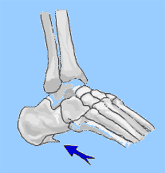
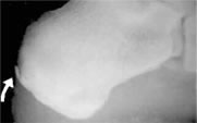
This
is a side (lateral) view of the heel bone, and demonstrates
a spur on the back (posterior) surface of the heel bone
(calcaneus).
This was asymptomatic (without
pain or other symptoms). A tight Achilles tendon can
cause this to form. It may be associated with pain at
the point were the achilles tendon inserts into the calcaneus.
|
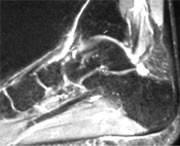
Below: Magnetic Resonance
Image (MRI) of Plantar Fasciitis showing a calcaneal spur

|

